Microscopic Improvements Make a Big Impact
Feb 26, 2021 — Atlanta, GA

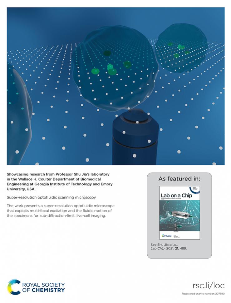
Illustration of super-resolution optofluidic scanning microscopy, which allows for imaging of living cells in flow. (Illustration Courtesy: Shu Jia)
By Zoe Elledge
For the first time, a microscopy system has been able to demonstrate super-resolution imaging of living cells in flow.
Walter H. Coulter Department of Biomedical Engineering Assistant Professor Shu Jia, along with his Laboratory for Systems Biophotonics, recently introduced their super-resolution optofluidic scanning microscopy system (OSM). It can view sub-diffraction-limit details of flowing cells and includes a high-quality microscope, a microfluidic system, and a micro lens array. These elements combine to create a grid of light spots that illuminate the sample inside a microfluidic channel.
Current microscopy technologies often sacrifice high-resolution images for a high throughput rate — the number of cells moving through the system to be analyzed. These systems need to stop the flow of cellular material in order to obtain a high-resolution image and therefore disturb the throughput rate. The flaws inherent in the current systems pose problems to researchers who need to analyze a large number of samples and want to take high-resolution images continuously. Jia’s new OSM system provides users the ability to do both.
“When you want to look at a cell, much of its organelles and structures are smaller than the conventional limit of the microscopes,” Jia said. “You want to have a higher resolution so that you can resolve finer structures. We’re trying to provide a system that can generate super-resolution images of the cells in flow so that you can learn more information from the cells and glean more biological insights.”
Jia and his team described their optofluidic scanning microscopy technology in the Royal Society of Chemistry journal Lab on a Chip. Their study appeared on the back cover of the third issue for 2021.
The OSM system illuminates the flowing sample in a pattern called multi-focal excitation, which provides super-resolution images of the sample and allows the team to extract even more information during analysis. Multi-focal excitation allows the system to take images without disrupting the flow of samples and makes it a revolutionary addition to the field of microscopy.
Another unique feature of the OSM is its platform accessibility, which is currently a topic of concern in the field of super-resolution microscopy. Jia’s lab created OSM to be compatible with various types of devices and samples so that its use can be broad and interdisciplinary.
“Just like a regular microscope, a lab can use it to image any sample it needs,” said Biagio Mandracchia, the paper’s first author and a postdoctoral fellow who works in Jia’s lab. “It offers a variety of opportunities for different disciplines and levels of research.”
Looking forward, OSM could be applied to fundamental biology studies, providing super-resolution images of large cellular populations and the individual organelles within a single cell. It could also be used to analyze tissue samples in biopsies. Jia said the technology could be used in preclinical and clinical studies, offering large amounts of diagnostic information faster.
“Our technique is simple, so we expect to see it used by physicians for obtaining diagnostics and analyzing samples, which will potentially have a large impact in both fundamental and clinical research,” he said.

Back cover of the third 2021 issue of the journal Lab on a Chip, featuring an illustration of Shu Jia's super-resolution optofluidic scanning microscopy.
Joshua Stewart
Communications Manager


